
Precision Diagnostics
3D CBCT Imaging & Digital X-Rays
We see structural details that traditional film x-rays miss. In-office 3D Cone Beam CT scans allow for safe, predictable dental surgery.
What is 3D Cone Beam CT Imaging?
Traditional dental X-rays provide a flat, two-dimensional picture. While useful, they cannot reveal the volume, density, or width of bones, nor do they map nerve coordinates or sinus locations with complete accuracy.
Cone Beam Computed Tomography (CBCT) is a medical scanning technology that rotates around your head, capturing hundreds of high-resolution digital exposures in a single 15-second sweep. Our computers then compile these exposures into a highly detailed3D anatomical model of your jaws, sinuses, and nerve pathways.
Safeguarding Your Oral Health
- Ultra-Low Radiation: Our Carestream digital systems emit up to 90% less radiation than traditional medical CT scans and film.
- Sub-Millimeter Precision: Essential for planning the exact angle and depth of dental implants to avoid major nerves.
- Immediate Diagnostic Output: Scan results populate on chairside monitors instantly, allowing Dr. Tafreshi to walk you through your anatomy.
Our Scanning Tech
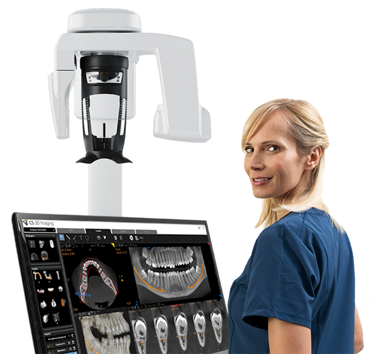
In-office low-dose Carestream dental 3D scanner system.
The Carestream CS 8200 3D Access Edition
Redefining diagnostic precision, the CS 8200 3D provides ultra-high-resolution images that reveal anatomical details hidden to traditional 2D X-rays.
Diagnose Hidden Issues
Detect microscopic fractures, nerve positions, and bone density variations.
Minimized Radiation
State-of-the-art dose management prioritizes patient safety without compromising clarity.
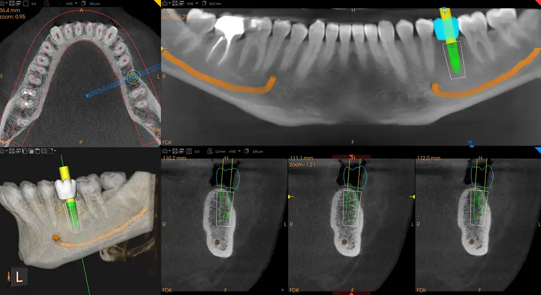
3D CBCT software planning for guided implant surgeries.
Clinical Benefits of In-Office 3D Imaging
1. Predictable Guided Implant Surgery
We compile your 3D bone maps with digital intraoral impressions to construct customsurgical guides. This guide ensures dental implants are seated in the absolute strongest bone columns, maximizing implant integration rates.
2. Safe Wisdom Teeth Extractions
Third molars are often located close to the inferior alveolar nerve canal. Our 3D scans show Dr. Tafreshi the exact spatial gap between the tooth root and the nerve, avoiding the risk of nerve irritation or numbness.
3. Root Canal Micro-Anatomy
Some teeth have hidden, curved, or accessory root canals that 2D x-rays fail to capture. Scans reveal these channels, preventing reinfection and root canal failures.
Frequently Asked Questions
Book Your Visit Today
Same-day appointments and free consultations available. Our team is ready to help.
Patient education
Keep your smile moving forward
Read practical treatment guides and oral-health answers from the AVF Dental Group team.

Zirconia Crown: Advantages and Disadvantages of Zirconia Crown
Is zirconia better than porcelain? How long do zirconia crowns last? A complete look at the strongest dental crown material available today.
Dental Veneers: Pros and Cons of Porcelain and Composite Veneers
Comparing porcelain, composite, and Lumineer veneers — advantages, disadvantages, candidacy, and cost considerations.
Wisdom Tooth Removal: When Is Wisdom Tooth Extraction Necessary?
Not every wisdom tooth needs to come out. Here is how dentists decide which third molars to remove and which to leave alone.